
Autopsy Documentation
External examination is photographed exhaustively and then the internal examination changes the body. Whatever was not photographed from the right angle is not in the record.
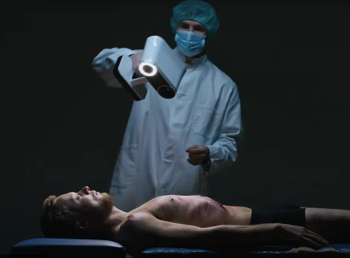
In a University of Toronto study of injury documentation, 11 simulated injuries on a live participant were recorded in 26 minutes with Artec Eva against 54 for photography and manual measurement — as colour 3D that can still be measured afterwards.
 No sensitive case details required.
How long does 3D injury documentation take?
In a University of Toronto study, documenting 11 simulated injuries on a live participant took 26 minutes with Artec Eva, against 54 for photography and manual measurement. Processing came afterwards, and the colour model could then be measured, including along curved skin.
 26 vs 54 minutes11 simulated injuries, Artec Eva vs photography (Toronto study).
 Colour and geometryLividity and injury appearance, with measurable form.
 ContactlessNo scale placed on the body, no camera set-up for each injury.
 ConsultableA second pathologist reviews the model, not a print.
Why the external record deserves better than a camera
Photography at autopsy is a compromise between time and coverage. Every injury is photographed with a scale, from the angles the photographer chose, under the lighting available; distances between injuries, the curvature of a wound on a limb, the true size of a pattern injury are all approximated from those frames afterwards.
The University of Toronto tested 3D scanning against photography on 11 simulated injuries marked on a live participant: 26 minutes with Artec Eva against 54 for photography and manual measurement, with processing done afterwards. The scan is measurable colour geometry, so a measurement not taken at the time — including one along curved skin — is taken later by anyone with the file. Artec Eva records the body; Artec Spider II records individual injuries at 0.05 mm.
For a medical examiner's office, the record is also the consultation: a second opinion is a file transfer, and the model can be shown in court where the court allows it.
The workflow, end to end
The whole body first, injuries second, before the internal examination.
 1. Capture the bodyWhole-body colour scan, anterior and posterior, in minutes.
 2. Capture injuriesClose-range scans of individual injuries at 0.05 mm.
 3. ProcessFuse in Artec Studio; register injury scans into the body model.
 4. MeasureDimensions, distances, curvatures — after the examination, from any angle.
 5. Archive and consultAttach to the case file; share with a second pathologist or the court.
Which scanner for this work
A body is captured with a fast handheld; injuries with a fine one. The Toronto study used the fast one, Artec Eva, alone.
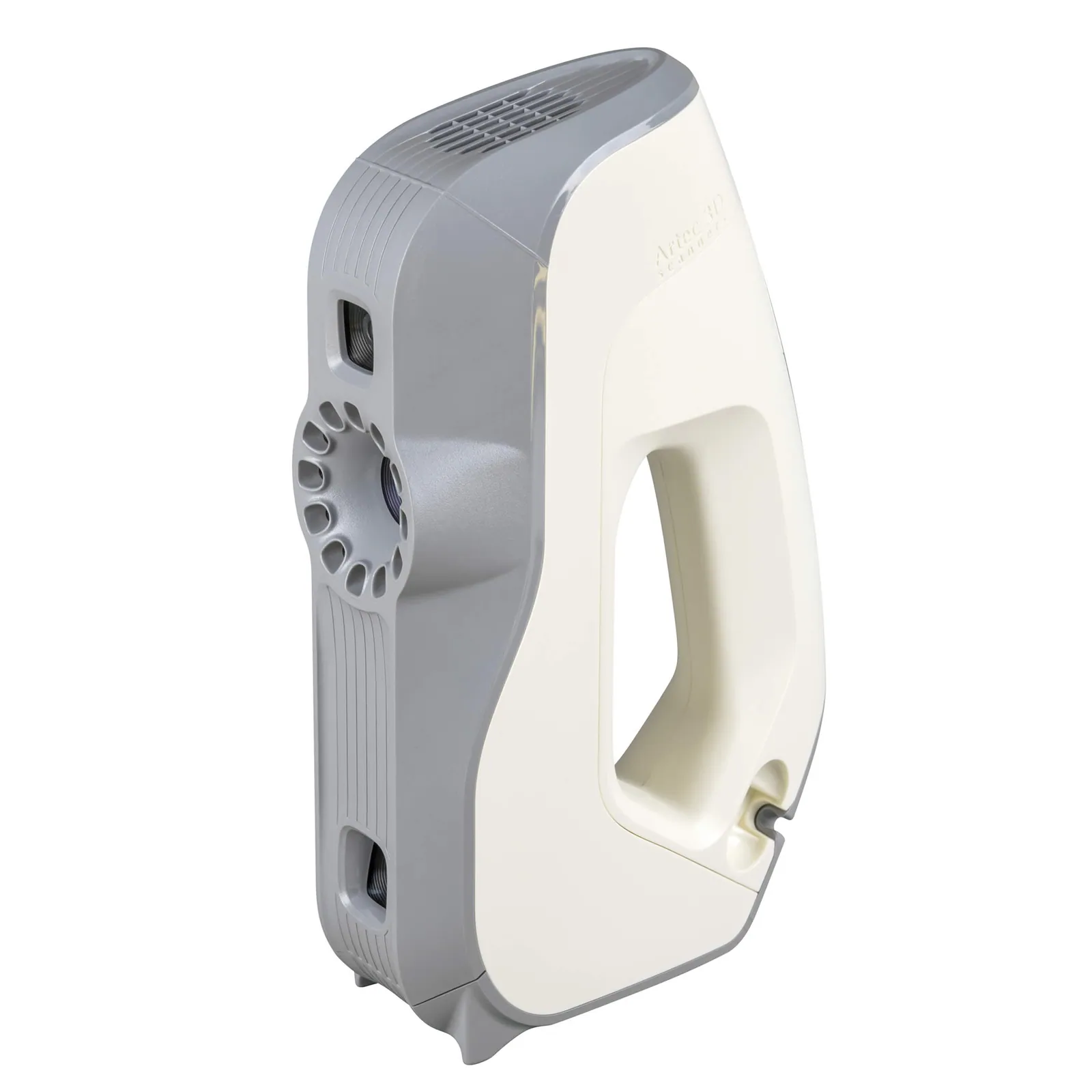
Artec Eva
- Best for
- Whole-body documentation
- 3D point accuracy
- Up to 0.1 mm
- 3D resolution
- Up to 0.2 mm
- Light source
- White structured light, full colour
- Portability
- Handheld, tethered to a laptop
- Toronto study
- 26 min for 11 simulated injuries, vs 54 for photography
Eva captures a body in colour, handheld and without contact, and it was the scanner in the Toronto study: 26 minutes for 11 simulated injuries on a live participant. Skin needs no preparation.
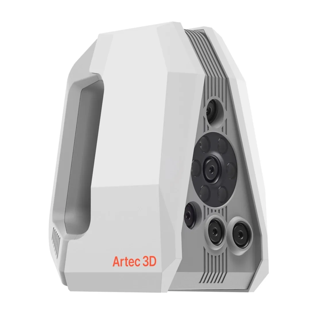
Artec Spider II
- Best for
- Individual injuries and pattern marks
- 3D point accuracy
- Up to 0.05 mm
- 3D resolution
- Up to 0.05 mm
- Light source
- Blue structured light, no targets on the surface
- Portability
- Handheld, tethered to a laptop
Spider II records a wound, a bite or a pattern injury at up to 0.05 mm accuracy and 0.05 mm resolution with colour, registered into the body model so its position is kept.
See external findings captured as measurable colour 3D, without contact — book a demo for your office.
Book a demoQuestions practitioners ask
Where does the 26-minute figure come from?
From a University of Toronto study comparing 3D scanning with forensic photography on a live participant marked with 11 simulated injuries, published as an Artec case study. Documenting them took 26 minutes with Artec Eva and 54 with photography and manual measurement; processing the model came afterwards.
Does the scan replace photography?
Not necessarily. Photography can be kept for specific purposes, with the scan adding measurable geometry alongside it; how the two are combined is a protocol decision for the office.
Can injuries be measured afterwards?
Yes, at any time, from any angle, including distances and curvatures that were not measured during the examination.
Is the body prepared or marked?
No. Capture is optical and contactless; no scales are placed and no markers applied.
Related applications
Tell us about the work you do
A forensic application specialist will come back with the device that fits it, the accuracy to expect at that scale, and how the data reaches your existing workflow.