
Forensic Odontology
Dental evidence is precise — arch form, tooth position, restorations, wear — and it is compared against records made by different people at different times.
3D scanning records dentition and bite injuries at 0.05 mm as measurable colour geometry, without an impression on the evidence or the tissue.
 No sensitive case details required.
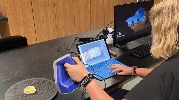
How is 3D scanning used in forensic odontology?
Dentition, dental casts and bite-mark injuries are scanned at up to 0.05 mm with colour; dental models are measured and compared for identification, bite injuries are preserved for review, and all are kept as a permanent record.
 0.05 mmArch, tooth position and restorations as measurable geometry.
 No impression materialNothing pressed onto an injury or fragile dentition.
 Model comparisonPost-mortem dentition against an ante-mortem cast, cast against cast.
 Permanent recordRemains released; the dentition remains as a file.
Why dental comparison is better done on models than on impressions
Forensic odontology compares shapes: a dentition against ante-mortem records, a suspected biter's teeth against an injury. Traditionally that means impressions and casts — material on evidence and distortion in the material. Bite mark analysis needs particular care: PCAST (2016) and NIST (2023) found it lacks a sufficient scientific foundation, not least because skin distorts the mark. A 3D record preserves the injury for review; it does not validate the comparison.
Artec Spider II records dentition, casts and injured tissue at up to 0.05 mm accuracy and 0.05 mm resolution with colour, without contact. Models are measured and compared in software; a bite injury on a body is recorded in position on the body scan. Artec Micro II records casts and individual teeth at up to 0.005 mm accuracy on an automated turntable for fine comparison.
The record outlasts the remains and the injury. It is also what a second odontologist reviews, and it can be shown in court where the court allows it.
The workflow, end to end
Record the injury, record the dentition, measure both as geometry.
- 1. Capture the injuryBite mark scanned in position on the body, at 0.05 mm with colour.
 2. Capture the dentitionTeeth, or a cast of them, scanned contactlessly.
 3. ProcessWatertight models in Artec Studio.
- 4. MeasureMeasure arch, spacing and tooth positions; any comparison with an injury is the odontologist's, within current guidelines and the limits NIST (2023) sets out.
- 5. ArchiveBoth records kept for review by another examiner.
Which scanner for this work
Injury and dentition with a handheld; casts and single teeth on the bench.
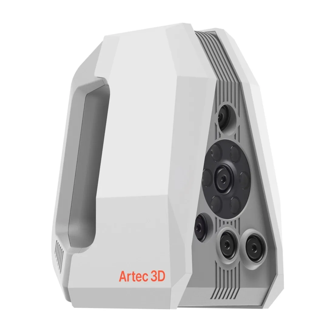
Artec Spider II
- Best for
- Bite injuries and dentition in place
- 3D point accuracy
- Up to 0.05 mm
- 3D resolution
- Up to 0.05 mm
- Light source
- Blue structured light, no targets on the surface
- Portability
- Handheld, tethered to a laptop
Spider II records an injury on skin, or dentition directly, at up to 0.05 mm accuracy and 0.05 mm resolution with colour. Nothing touches the tissue or the teeth.
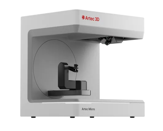
Artec Micro II
- Best for
- Casts and individual teeth
- 3D point accuracy
- Up to 0.005 mm
- 3D resolution
- Up to 0.04 mm
- Capture
- Desktop, fully automated turntable
- Object size
- Up to 200 × 200 × 150 mm
Micro II records a dental cast or a single tooth at up to 0.005 mm on an automated turntable — a metrology-grade record for comparison and for restorations.
See dentition and bite injuries recorded as measurable geometry — book a demo for your odontology team.
Book a demoQuestions practitioners ask
Can teeth be scanned directly in the mouth?
Post-mortem dentition is scanned directly with Spider II where access allows; otherwise a cast is scanned. Intra-oral scanning of the living is a dental-scanner task.
How is a bite mark compared?
Both models can be overlaid in Artec Studio or exported to your analysis software, and arch form, spacing and tooth positions measured. The interpretation is the odontologist's, within the limits PCAST (2016) and NIST (2023) set out: skin distorts a bite mark, and the comparison lacks a sufficient scientific foundation.
Can the scan be compared with ante-mortem dental records?
Yes. Restorations, missing teeth, rotations and arch form are measured on the post-mortem model and set against charts, radiographs or a scanned cast from the dentist, alongside the post-mortem radiographs the comparison relies on; the comparison is repeatable by a second odontologist.
How does it help identification?
The post-mortem dentition becomes a permanent, measurable file that is compared against ante-mortem records and kept after the remains are released.
Tell us about the work you do
A forensic application specialist will come back with the device that fits it, the accuracy to expect at that scale, and how the data reaches your existing workflow.