
Human remains and pathology in 3D
Remains are handled once if you are careful, and every handling costs something. A 3D record lets the examination continue on the model: measure a fracture margin, revisit a wound, share a finding with a specialist on another continent.
Eight areas below, from field recovery through identification.
 No sensitive case details required.
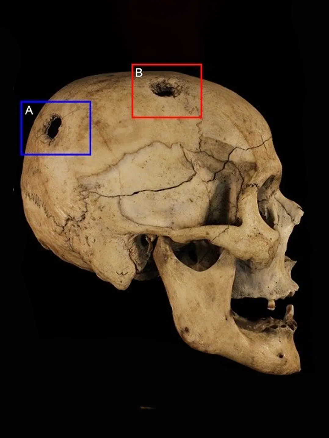
 ContactlessNo contact, no coating. Safe for fragile bone and for the examiner.
 Colour with geometryDiscolouration and staining recorded alongside shape, in one dataset.
 Measure afterwardsAny dimension, any time, without recalling the remains.
 Share for reviewA specialist opinion without shipping anything anywhere.
What are you documenting?
Field recovery, laboratory examination and identification each need a different capture scale.
Why the record has to be measurable, not photographic
A photograph of a fracture shows that it is there. It does not give the margin, the angle or the relationship to the bone around it — and those are the measurements an opinion rests on. Once remains are released, buried or repatriated, a photograph cannot answer a question nobody thought to ask.
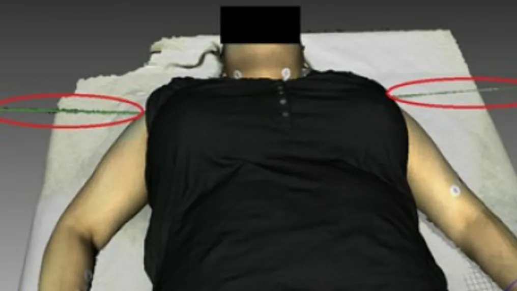
In a University of Toronto study, documenting 11 simulated injuries on a live participant took 26 minutes with Artec Eva against 54 for photography and manual measurement (processing done afterwards), and the model could be measured along curved skin. The capture itself is contactless, so it adds little to the handling the remains already receive.
Measured, not asserted
What the model supports afterwards

Measurement on demand: Fracture length, wound depth, angle between surfaces — taken from the archive.

Several specialists, one dataset: Anthropologist, pathologist and odontologist work from the same capture.
Remote review: A second opinion without transporting remains or waiting for a visit.

After release: The record outlives the availability of the remains themselves.
See remains and injuries documented at research resolution, without contact — book a demo for your department.
Book a demo“Artec has truly changed the face of forensic anthropology with Space Spider, Leo, and Ray. I’ve integrated Artec’s solutions into my daily field work as well as my regular and special classes for investigators, law enforcement officers, and college students, and the results have been nothing short of spectacular.”
Questions that come up first
Does scanning add handling to the examination?
Very little. Capture is optical and contactless: no targets are placed on the remains and nothing is applied to the surface; an element is turned only to record its other side. It can be done at the point in the examination where the remains are already exposed.
Which scanner for bone detail?
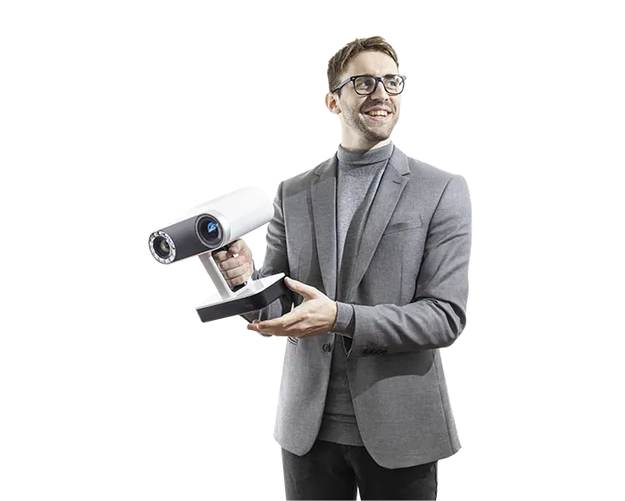
Artec Spider II for trauma margins, dental detail and small elements, at 0.05 mm accuracy. Artec Leo or Eva for whole bodies and articulated skeletons, where field of view matters more than the last fraction of a millimetre.
Can the model be used for identification work?
A skull scan is a normal starting point for facial approximation, and dental scans support comparison against ante-mortem records. What the model does is preserve the geometry those methods depend on; the methods themselves stay yours.
Is this used in teaching as well as casework?
Yes — teaching collections are one of the established uses, because a scanned element can be handled by a class without the original leaving its store. Dr Dennis Dirkmaat, then teaching at Mercyhurst University, reported that his students picked up the scanning process quickly.
Other areas of forensic 3D work
Tell us about the work you do
A forensic application specialist will come back with the device that fits it, the accuracy to expect, and how the data reaches your existing case workflow.