
Forensic Anthropology
The biological profile — age, sex, stature, ancestry — is built from morphology and measurements on bone that is fragile, often contested and frequently returned before the questions stop.
3D scanning records each element at up to 0.05 mm with colour, so the osteometrics, the taphonomy and the second opinions are taken from the model.
 No sensitive case details required.
What does 3D scanning give a forensic anthropologist?
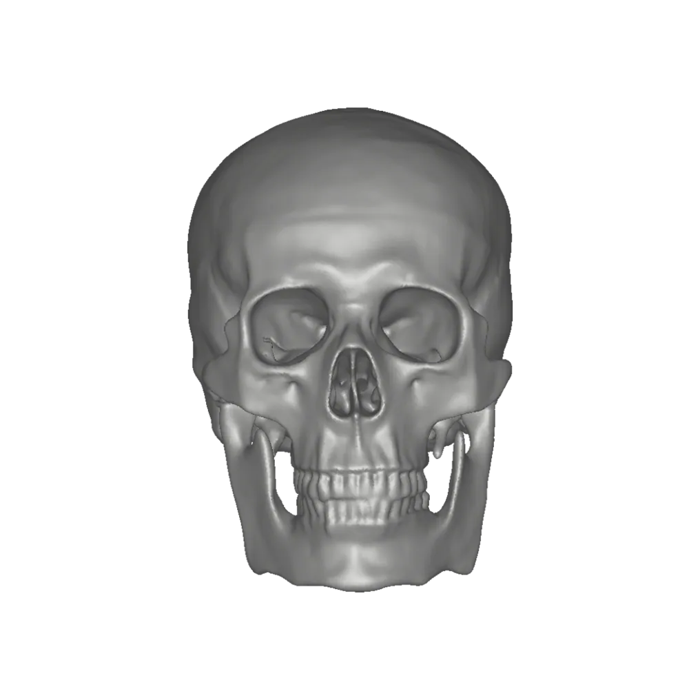
A colour model of each element at up to 0.05 mm on which osteometric landmarks are placed, weathering and staining are read, trauma is assessed and teaching continues — after the remains have been released.
 Osteometrics on the modelLandmarks placed and inter-landmark distances measured digitally, repeatably.
 Taphonomy in colourWeathering stages, staining and root etching recorded with the geometry.
 0.05 mm for traumaKerf walls and fracture margins at the resolution the assessment needs.
 Teaching and consultationOne specimen, every student; a colleague abroad measures the same element.
The biological profile, built on geometry that outlasts the remains
Sex is estimated from pelvic and cranial morphology and from measurements; stature from long-bone lengths; adult age from the morphology of the pubic symphysis and auricular surface; ancestry from cranial form. Every one of them is a measurement or a shape judgement on an element that is handled to make it — and handled again when a second anthropologist checks it. Photographs record what one lighting angle showed; callipers record what one hand measured.
A model at up to 0.05 mm from Artec Spider II carries the landmarks and the morphology together, in colour, so taphonomic change — weathering stages, root etching, staining, carnivore damage — is read from the same file as the metrics. Trauma assessment continues on the same model, and the element goes back into its box. For teaching, a scanned reference collection puts one specimen in front of every student without wear on the original.
In the field the discipline meets recovery work: remains recorded in situ before they are lifted, on the terms described under clandestine graves. Dr Dennis Dirkmaat, while at Mercyhurst University, used Artec scanners at outdoor death scenes to record the bones and the scene around them.
The workflow, end to end
Field first if the remains are still in the ground; bench for every element after.
 1. Record in situRemains and context captured where they lie, before recovery.
- 2. Scan each elementOn the bench at 0.05 mm with colour — cranium, pelvis, long bones, dentition.
- 3. Place landmarksOsteometric landmarks on the model; inter-landmark distances measured digitally.
 4. Read taphonomy and traumaWeathering, staining and defects assessed on the colour model, sectioned where needed.
 5. Archive, teach, consultRemains released; the model stays for the report, the course and the second opinion.
Which scanner for this work
One scanner for the bench, one for teeth and fragments. Whole-skeleton layout capture is covered under human skeletal remains.
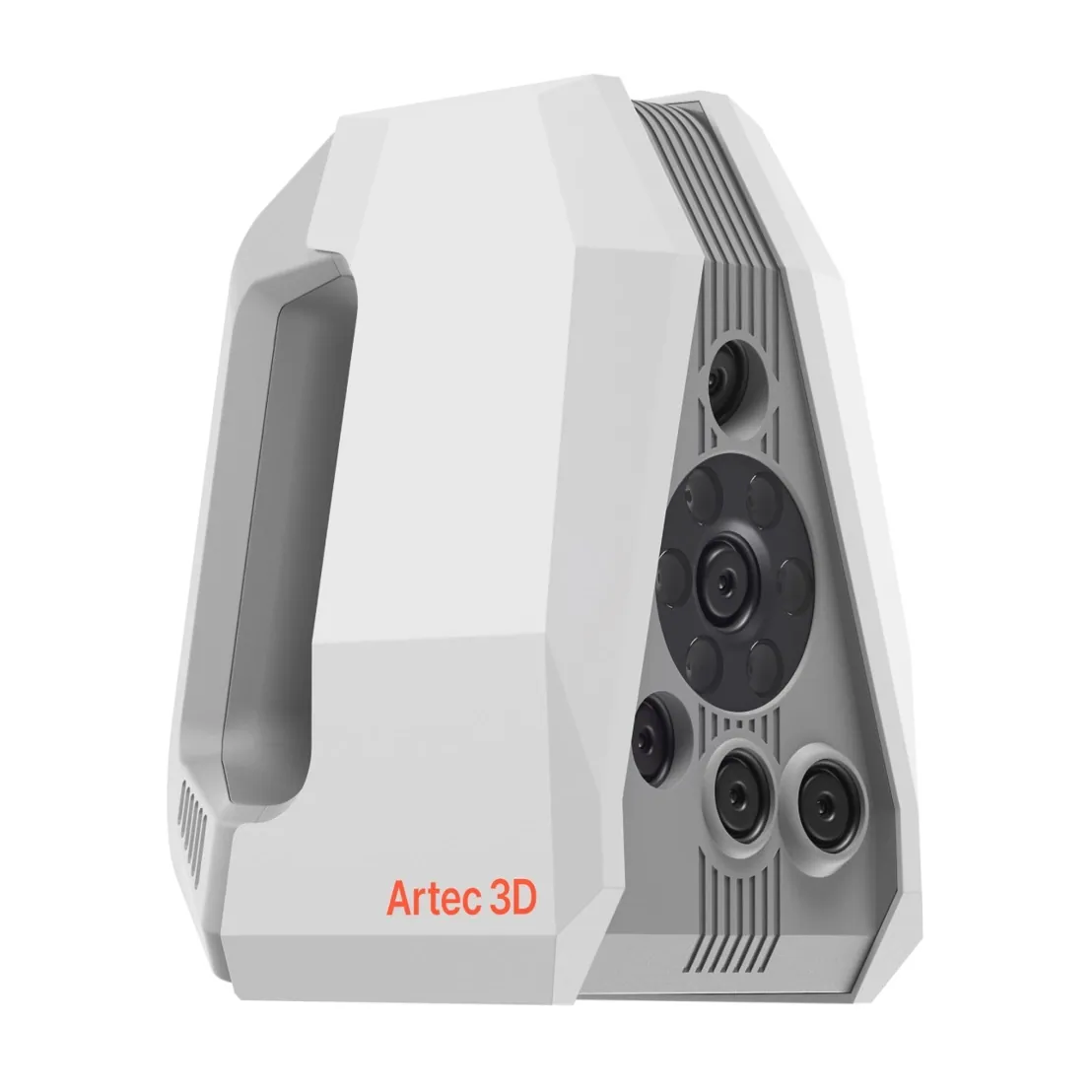
Artec Spider II
- Best for
- Elements on the bench — landmarks, taphonomy, trauma
- 3D point accuracy
- Up to 0.05 mm
- 3D resolution
- Up to 0.05 mm
- Light source
- Blue structured light, no targets on the surface
- Portability
- Handheld, tethered to a laptop
Spider II records bone at up to 0.05 mm accuracy and 0.05 mm resolution with colour, without contact. Osteometric landmarks are placed on geometry rather than felt for with callipers; weathering and staining come through in the texture.
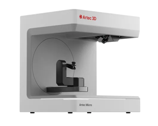
Artec Micro II
- Best for
- Teeth, small bones and fragments
- 3D point accuracy
- Up to 0.005 mm
- 3D resolution
- Up to 0.04 mm
- Capture
- Desktop, fully automated turntable
- Object size
- Up to 200 × 200 × 150 mm
Micro II records small elements at up to 0.005 mm on an automated turntable — a metrology-grade record for dentition, hand and foot bones and fragments, for research as well as casework.
“Artec has truly changed the face of forensic anthropology with Space Spider, Leo, and Ray. I’ve integrated Artec’s solutions into my daily field work as well as my regular and special classes for investigators, law enforcement officers, and college students, and the results have been nothing short of spectacular.”
See osteometric landmarks placed and taphonomy read on a scanned element at 0.05 mm — book a demo for your department.
Book a demoQuestions practitioners ask
Are osteometric measurements on the model equivalent to callipers?
Inter-landmark distances are measured on geometry stated at 0.05 mm accuracy, and the landmark placement is recorded, so a second anthropologist repeats the same measurement on the same file. Validate against your calliper protocol on reference material before switching.
Does the scan capture taphonomic change?
Yes — colour texture is captured with the geometry, so weathering stages, staining, root etching and carnivore damage are in the record alongside the shape.
Can the models be used for teaching?
That is one of the main uses. A scanned reference collection is measured by every student on screen, printed as replicas from watertight STL, and never wears the original.
How does this connect to field recovery?
Remains are recorded in situ with a wireless handheld before they are lifted; the method is described under clandestine graves. The bench work then continues on the same case.
Tell us about the work you do
A forensic application specialist will come back with the device that fits it, the accuracy to expect at that scale, and how the data reaches your existing workflow.