Medical Examiners / Pathologists

An office answers for a protocol, a caseload and a record that will be read by a court years later — often after the pathologist who made it has moved on.

3D documentation changes the protocol once and then pays on every case: less time than photography, measurements taken afterwards from any angle, and a second opinion that is a file transfer.

No sensitive case details required.

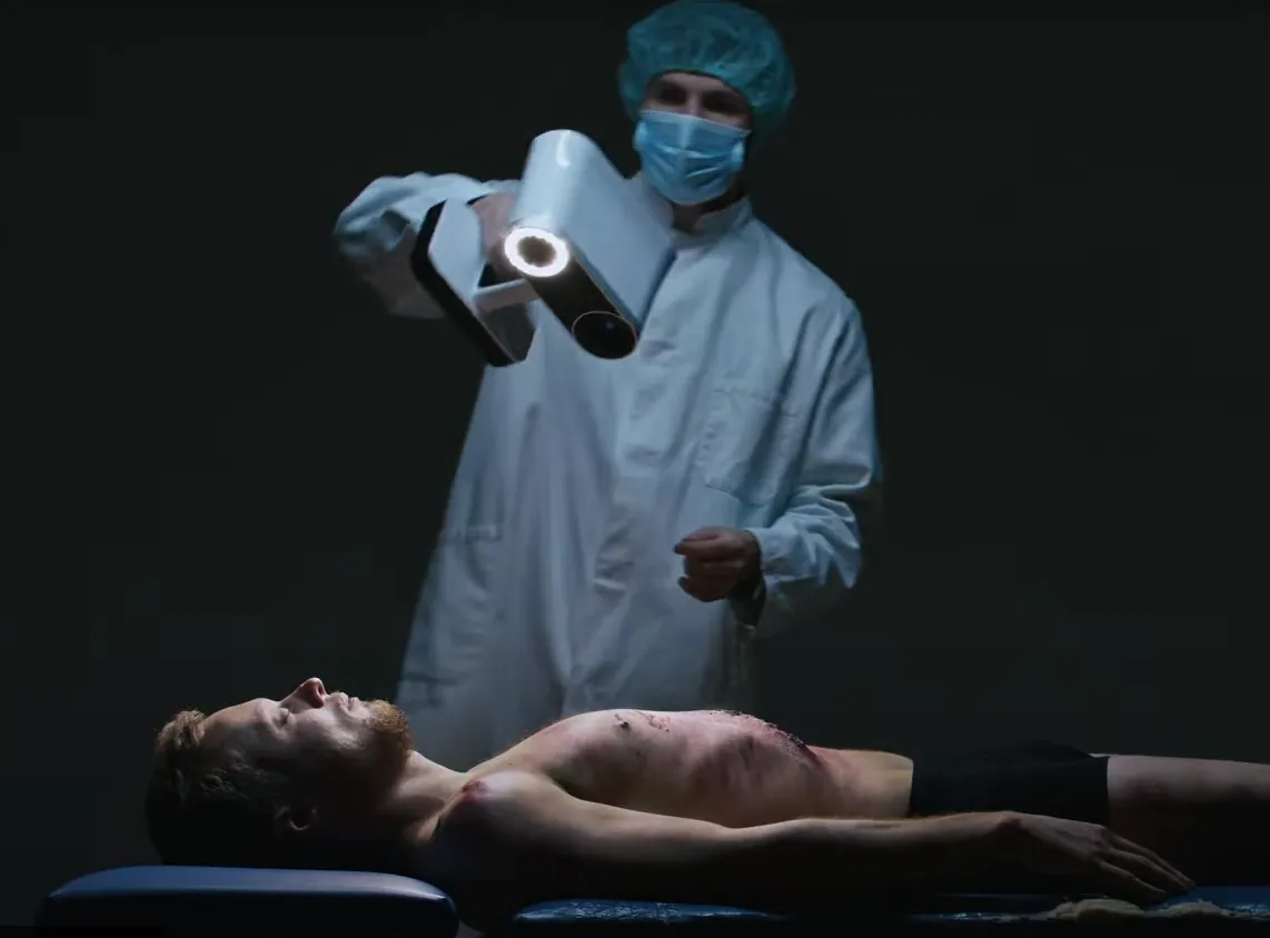
3D scanning a dead body with Artec Leo
  • Less time per caseA full external record in less than half the photography time in the Toronto comparison.
  • Measure after the factDistances and dimensions not taken at the time, taken later from the model.
  • Consultation by fileA second pathologist reviews the model, not a print.
  • Court without the photographsA model or a print, shown without images a jury struggles with.

What changes for the office

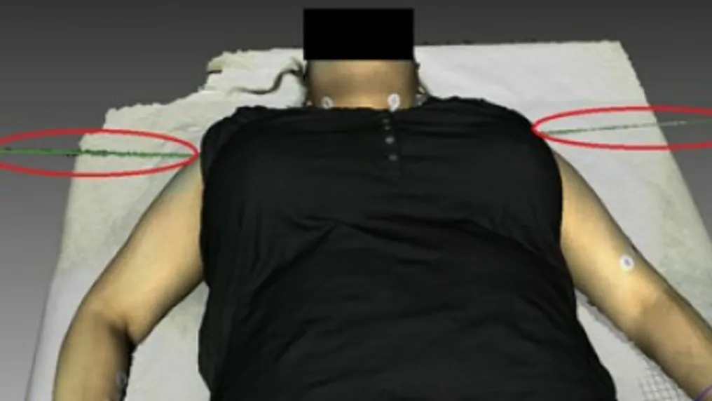
3D scan of a body with measurements marked on the model
  • The protocol changes once: The scan takes a fixed place in the external examination; photography becomes targeted rather than exhaustive.

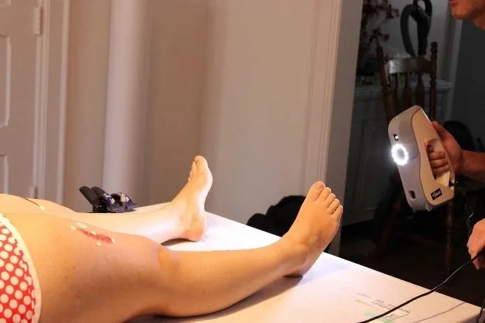
  • Injuries at 0.05 mm: Wounds, bites and pattern injuries recorded as measurable geometry, registered into the body model.

  • Retention that answers questions: The record outlives the examination, the exhumation question and the retirement of the pathologist.

  • A record the court reads: A model, a section or a print — presented without the photographs a jury finds hard to read.

Protocol, caseload, consultation, court — what an office is accountable for

Adopting 3D documentation is a protocol decision before it is a device one. Where the scan sits in the external examination, what photography is still taken and for what, who processes and who signs — those are written once. The procedure itself is described under autopsy documentation; wound documentation and forensic odontology cover the close-range work.

On caseload the arithmetic is the University of Toronto comparison: a full external record in 26 minutes against 54 for photography, with submillimetre colour geometry the photographs could not give. Over an office's annual caseload that is not a saving at the margin.

The record then does the office's later work. A consultation with a specialist elsewhere is a file transfer; a question raised at trial is a measurement on the model; a case reopened years later is answered from the archive. Retention follows the office's existing rules for imaging, sized for larger files.

Peers already doing this

The figures are the customers' own, from their published case studies.

Which scanner for this work

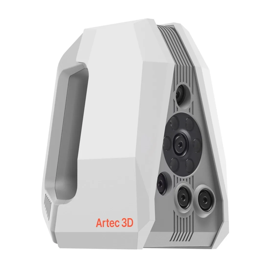
The combination the Toronto study ran: a fast handheld for the body, a fine one for the injuries.

Artec Eva 3D scanner

Artec Eva

Best for
Whole-body documentation
3D point accuracy
Up to 0.1 mm
3D resolution
Up to 0.2 mm
Light source
White structured light, full colour
Portability
Handheld, tethered to a laptop

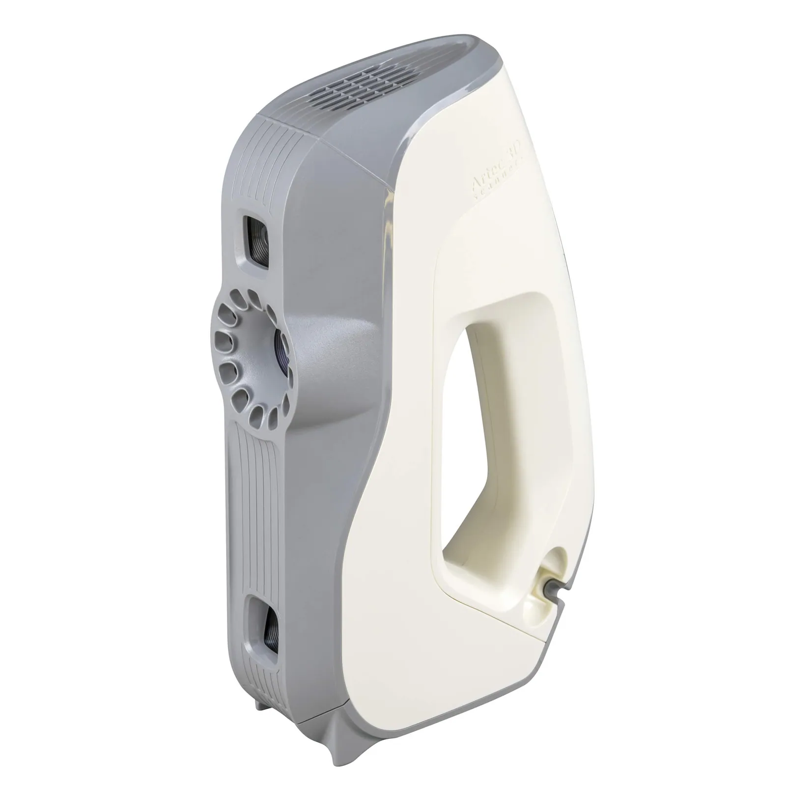
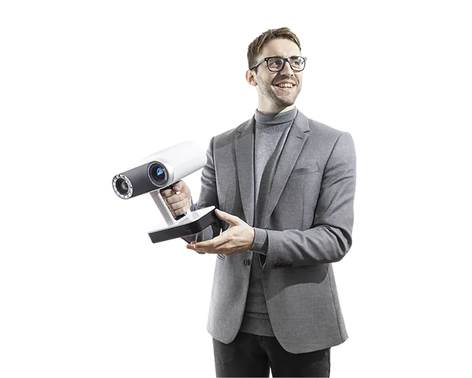
Eva captures a body in colour in minutes, handheld and contactless — the speed behind the 26-minute figure.

Artec Spider II 3D scanner

Artec Spider II

Best for
Wounds, bites and pattern injuries
3D point accuracy
Up to 0.05 mm
3D resolution
Up to 0.1 mm
Light source
Blue structured light, no targets on the surface
Portability
Handheld, tethered to a laptop

Up to 0.05 mm accuracy and 0.1 mm resolution with colour, registered into the body model so each injury keeps its position.

See what changes for an office that documents the external examination in 3D — book a demo.

Book a demo

Questions practitioners ask

How does the protocol change?

The scan takes a fixed place in the external examination — typically anterior and posterior before the internal examination — and photography becomes targeted: close-up appearance and alternative-light imaging rather than exhaustive coverage.

What does it save per case?

In the University of Toronto comparison, 26 minutes against 54 for a full external record. The rest of the saving is downstream: measurements taken later instead of a re-examination, and consultation by file.

How is retention handled?

As for imaging, sized for larger files: raw scans retained, processing documented, exports hashed. Scan projects can run to gigabytes per case.

Who processes the scans?

Autopilot processing in Artec Studio is run by a technician; the pathologist reviews and measures on the finished model.

Talk to a forensics expert

Tell us about the work you do

A forensic application specialist will come back with the device that fits it, the accuracy to expect at that scale, and how the data reaches your existing workflow.

No sensitive case details required.