Blood spatter analysis: how bloodstain patterns are read
Blood spatter analysis, properly called bloodstain pattern analysis, reads the size, shape and position of bloodstains to work out where blood came from, what set it in motion and in what order.
Bloodstain pattern analysis sorts stains into passive, transfer and spatter patterns, then uses their shape and position to reconstruct a bloodshed event.
A spatter stain's angle of impact is the arcsine of its width divided by its length; stains from one event converge on the area where the blood source was.
In the largest black-box study of the field (75 analysts, 192 patterns), 11.2% of analysts' responses on samples of known cause were wrong. The method is only as good as the measurements behind it.
A 3D scan keeps the stains' shapes and positions measurable after the scene is cleaned, so the analysis can be checked and repeated.
Blood spatter analysis is the examination of bloodstains at a scene to reconstruct what happened there: where a bleeding person or blood source was, what kind of force acted on the blood, how people moved and in what order things occurred. Analysts call the discipline bloodstain pattern analysis (BPA), because spatter is only one of the patterns they read.
This guide covers the pattern types, the physics that shapes them, the arithmetic behind the angle of impact and the area of origin, how patterns are documented, and how much weight the conclusions can bear.
What is blood spatter analysis?
Blood spatter analysis, or bloodstain pattern analysis, is a forensic discipline that examines the size, shape, distribution and location of bloodstains to infer the events that produced them. It treats each stain as the end of a physical process (a drop that fell, was flung or was transferred) and reasons back from the stain to that process.
From a well-documented pattern, an analyst can often establish:
where the blood source was, and roughly how high above the floor
the mechanism: a drip, a blow, a gunshot, a cut artery or blood breathed out
the direction a person or object moved, and sometimes how fast
a minimum number of blows struck with a bloodied weapon
whether a body or an object was moved after the bloodshed
the order of events, where one pattern lies on top of another
What it cannot do is say whose blood it is. That is DNA analysis. The two work together, and a pattern is documented before it is sampled, because swabbing changes it.
Blood spatter or blood splatter?
Spatter is the term analysts use, in their standards and in court; splatter is the everyday word and the more common search. In conversation they mean the same thing, but a report uses spatter, and only for stains from drops that a force sent through the air. A drop that fell under its own weight, or a smear on a wall, is a bloodstain but not spatter.
How blood behaves when it leaves the body
Every pattern comes from a few physical properties of blood. Blood is about 55% plasma, with red cells, white cells and platelets suspended in it, and it behaves as a shear-thinning, non-Newtonian fluid: the faster it is forced to move, the more easily it flows. In flight, surface tension pulls a drop into a sphere. Real drops do not have the teardrop shape of cartoons.
What happens on impact depends mostly on two things: the angle the drop arrives at and the surface it lands on. A drop striking a smooth, hard surface at 90° leaves a near-circular stain, and the shallower the angle, the longer the ellipse. A rough or absorbent surface such as carpet, raw wood or cotton breaks the edge of the drop into spines and satellites at almost any angle, which is why the surface itself is part of the evidence.
Analysts read four features on a single stain:
Parent stain: the main ellipse left by the drop itself. Its width and length give the angle of impact.
Spines: short points on the leading edge, more pronounced on the side the drop was travelling towards.
Satellite stains: small drops that broke away on impact and landed beyond the parent stain.
Tail: the tapering end that points in the direction of travel. It is left out when the length is measured.
Drop size tells you less than it seems to. The force, the volume of blood available and the surface all change it, which is one reason the old velocity categories described below were abandoned.
The three families of bloodstain patterns
Analysts classify stains by the mechanism that made them. A widely taught scheme has three families: passive stains, transfer stains and spatter. The classification by Stuart James, Paul Kish and Paulette Sutton counts transfer stains among the passive ones and adds altered stains, for stains that changed after they formed. The AAFS Standards Board's terminology, ASB Technical Report 033, defines the individual patterns.
PassiveGravity
TransferContact
SpatterProjection
Formed by
Blood falling or flowing under its own weight
A bloodied surface touching another
Drops sent through the air by a force
Typical patterns
Drips, drip trails, flows, pools
Swipes and hand, shoe or fabric impressions
Impact and gunshot spatter, cast-off, arterial, expirated
What it can show
Where a source stood or moved; whether a body was moved
What touched what, and in which direction it moved
The mechanism, the area of origin, a minimum number of blows
Altered stains (clotted, dried, diluted, wiped or cleaned) and voids can occur in any family.
Passive stains: drips, flows and pools
Passive stains form under gravity alone. A drip stain is a single drop that fell; a drip trail is a series of them left by a moving source, and its spacing hints at speed, with drops close together suggesting slow movement. A flow pattern is blood running across a surface. A flow that runs uphill in the position a body was found in shows the body was moved. A pool forms where a source stays still long enough for blood to collect, and often marks where a victim ended up.
Transfer stains: swipes and wipes
A transfer stain is left when a bloodied surface touches another one. A swipe carries blood onto a clean surface, as when a bloodied hand is drawn across a wall. A wipe is the reverse: something moving through blood that was already there and disturbing it, which is why the ASB terminology classes a wipe as an altered stain rather than a transfer. The feathered edge of a swipe usually thins out in the direction of movement. Pattern transfers, such as a shoe sole, a palm or the weave of a sleeve, can be compared with the object that made them.
Spatter: impact, cast-off, arterial and expirated
Spatter is blood that travelled through the air as drops, and it is grouped by what set it in motion:
Impact spatter: a force strikes liquid blood, as in a blow to a bleeding wound. The drops radiate from the point of impact, which is what makes an area of origin calculable.
Gunshot spatter: back spatter flies from the entrance wound back towards the firearm; forward spatter leaves with the bullet through an exit wound. Forward spatter usually holds more drops than back spatter, and either can include a fine mist; both are easy to miss, and the finest drops can be mistaken for expirated blood. Placing them is part of shooting scene reconstruction.
Cast-off: blood flung from a bloodied object as it swings, typically as linear trails on a ceiling or wall. The first blow usually meets no exposed blood, so it rarely leaves cast-off; the number of trails plus one is therefore a common, though contested, estimate of the minimum number of blows. A cessation pattern, often called cessation cast-off, is thrown when a swinging object stops abruptly.
Arterial pattern: blood pumped from a breached artery, in arcs or zigzags that rise and fall with the heartbeat. It shows the person was alive when the pattern formed.
Expirated spatter: blood breathed, coughed or sneezed out of the nose, mouth or an airway wound. It is usually fine and can carry tiny air bubbles, which separate it from impact spatter of a similar size.
Altered stains and voids
Blood changes after it lands. It clots within minutes, dries from the edge inwards, darkens from red to brown, and can be diluted, smeared or cleaned up. An analyst reads those changes as time and activity: clotted blood under fresh drops means bleeding went on; diluted stains point to water or cleaning. Cleaned blood is rarely gone. Luminol, which reacts with the iron in haemoglobin, makes faint traces glow in the dark.
A void is a gap in an otherwise continuous pattern, where a person or object stood between the source and the surface. A void with nothing in it says something was moved, and the object, if it is found, carries the matching stains.
Why "low, medium and high velocity" spatter is no longer used
Older textbooks sort spatter by speed: low-velocity stains of 4 mm and larger from drips, medium-velocity stains of 1 to 4 mm from blows, and high-velocity stains under 1 mm from gunshots, with the blow on the blood source put at up to 1.5 m/s, 1.5 to 7.5 m/s and 30 m/s or more.
The scheme was dropped because it confuses cause with size. The velocity described the blow that struck the blood source, not the speed of the drops; one blow can throw drops of every size; and a cough produces a mist as fine as a gunshot's. Calling a stain high velocity therefore implied a gunshot where the pattern could not show one, the kind of overreach behind several disputed cases. Current terminology names the mechanism the evidence supports, or simply describes the stain.
How to calculate the angle of impact
The angle of impact is the angle between a drop's flight path and the surface it struck, and it comes straight from the shape of the stain: the sine of the angle equals the stain's width divided by its length.
Angle of impact = arcsin (width ÷ length)
Choose a well-formed stain with a clear ellipse, not one distorted by a rough surface or a second drop.
Measure the width across the widest point and the length along the long axis, leaving the tail out.
Divide the width by the length and take the arcsine.
For a stain 4.0 mm wide and 8.0 mm long, width ÷ length = 0.50, and arcsin 0.50 = 30°. A round stain, where width equals length, gives 90°: the drop came straight down.
Small measuring errors matter. Reading that 4 mm width as 4.1 mm moves the angle to almost 31°, and on a stain 2 mm wide and 4 mm long the same 0.1 mm moves it by close to 2°. Those degrees add up when dozens of stains are projected back to an origin, which is why analysts measure many stains, fit ellipses rather than judging them by eye, and measure on the surface rather than on a photograph taken at a slant.
The stain's long axis also gives its direction across the surface, called its directionality, and the tail says which way along that axis the drop was moving.
Angle of impact = arcsin (width ÷ length). A stain 4.0 mm by 8.0 mm gives 30°.
Finding where the blood came from
Stains from one impact radiate from a single source, so their geometry can be run backwards. It takes two steps: the area of convergence on the surface, then the area of origin in the room.
Area of convergence: draw each stain's long axis back across the surface. The lines meet in a small area, which is where the source was in plan, in two dimensions.
Area of origin: rise from that area at each stain's angle of impact. The height is the distance from the stain to the convergence multiplied by the tangent of its angle: h = d × tan α.
A stain 80 cm from the area of convergence that struck the floor at 30° puts the source about 80 × tan 30° ≈ 46 cm above the floor: close to the floor, far below where a standing person's head would be. Repeating the calculation across many stains turns a point into an area, and the area is what gets reported.
Long axes meet at the area of convergence; the angles of impact lift it to the area of origin.
Three methods do this work:
Stringing: a string from each stain at its angle of impact, run back until the strings cross. It is physical and easy to show, but slow, and it ends when the strings come down.
The tangent method: the same geometry, calculated for one stain at a time.
Software: programs such as HemoSpat, BackTrack and FARO Zone 3D compute the area of origin from measured stains, working from photographs or from a 3D model of the scene.
All three treat flight paths as straight lines. Real drops fall under gravity and slow down in the air, so they arrive steeper than they left, and straight-line methods tend to place the origin higher than it was. That is why the area of origin is reported as a range, and why better stain measurements allow an honestly narrower one.
How a bloodstain pattern is documented
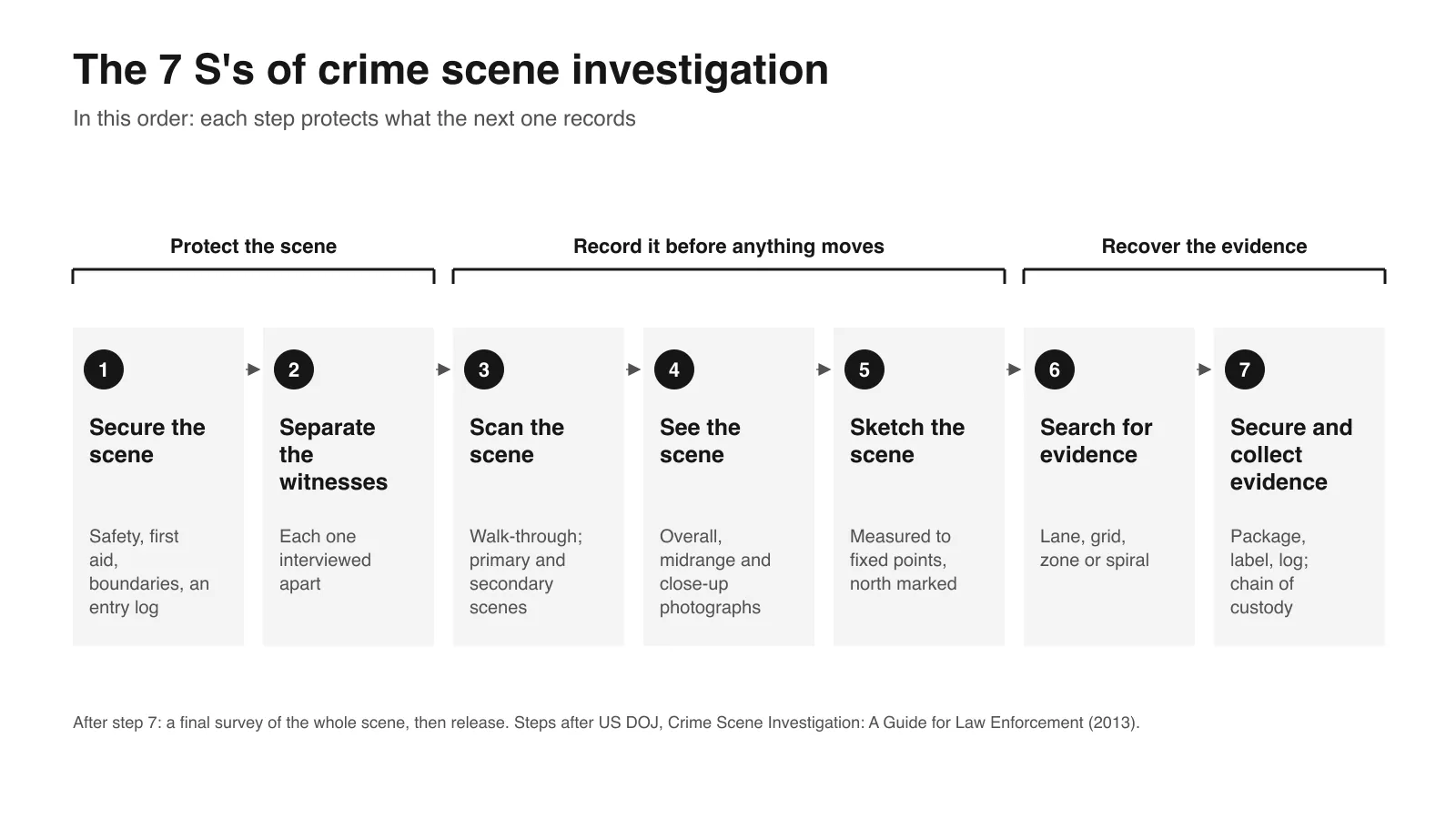
An analysis can be no better than its record of the scene, and most of the work happens before anyone interprets anything. A typical sequence:
Overall photographs and a scene record before anything is touched, then mid-range and close-up photographs of each pattern.
A scale in every close-up, placed in the plane of the stain, with the camera square to the surface.
Labelling and measuring: stains of interest are numbered, often against a grid or reference line taped across the surface, and each one's position, width, length and direction recorded.
Sampling for DNA, only once the pattern is fully documented.
Measurement of the room itself, so every stain can be placed in three dimensions.
Each step has a known weakness. A photograph flattens the surface and distorts exactly the width-to-length ratio the angle depends on, and a scale corrects for magnification but not for a camera a few degrees off square. Measuring stains by hand is slow, and once the scene is released and cleaned, nothing that was missed can be measured again.
That is the part 3D scanning has changed. A scan records each stain's shape together with the surface it sits on, and places it in a measured model of the room, so the angle of impact and the area of origin can be recalculated later by the same analyst, a reviewer or the other side's expert. 3D scanning for bloodstain pattern analysis covers the method in detail, and the same capture serves 3D crime scene reconstruction.
Tape and photographsEach stain by hand
StringingStrings in the room
3D scanStains and room in one model
Time at the scene
Long: every stain measured in place
Hours for a large pattern
Minutes of capture per area
Re-measurable after release
Only from the photographs
No: the strings come down
Yes: the scan is the record
Area of origin
Calculated separately
Physical and approximate
Computed from the model, any time
What reaches the court
Photographs and a table of figures
Photographs of the strings
A measurable 3D model
Times depend on the size of the pattern and the scene; what differs is the order of magnitude.
Used for this in practice
Artec Spider II
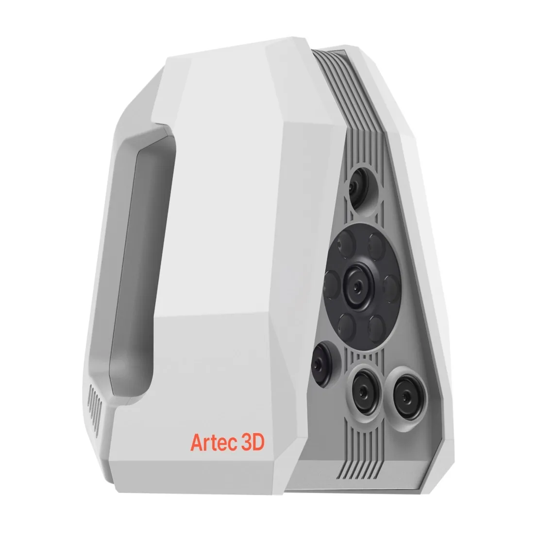
A handheld structured-light scanner that records a stain and the surface under it together, so its width and length are measured on the true surface rather than a flattened photograph, with nothing touching the stain. Paired with an Artec Ray II dome scan of the room, every pattern sits in one coordinate system.
Blood spatter analysis is not debunked, but its limits are documented, and they are wider than television suggests. The physics of how drops form, fly and strike is well understood. The weak point is the human interpretation of complex patterns.
The 2009 National Academy of Sciences report concluded that the uncertainties in bloodstain pattern analysis are enormous and that many experts' opinions were more subjective than scientific. In 2021, an NIJ-funded black-box study tested 75 practising analysts on 192 patterns chosen to represent real casework. On samples whose cause was known, 11.2% of responses were wrong, and across the study 7.8% of responses contradicted those of other analysts, although the consensus of the group was seldom wrong.
The authors found that semantic differences, analysts using the same terms for different things, contributed to the errors alongside genuinely contradictory interpretations, and called for better standards for terminology and classification. The other recurring problems are thin documentation, ambiguous patterns with few stains, and context: an analyst who knows the investigators' theory is more likely to see it.
In England and Wales, the Forensic Science Regulator publishes BPA guidance under its Code of Practice. In the US, the field has responded with consensus standards from the AAFS Standards Board:
ASB Technical Report 033: terms and definitions (2017)
ANSI/ASB Standard 030: quality assurance programmes (2019)
ANSI/ASB Standard 072: validation of procedures (2019)
ANSI/ASB Standard 031: report writing (2020)
ANSI/ASB Standard 032: training programmes (2020)
What makes a conclusion defensible is the same everywhere: measurements anyone can check, a documented method, and an opinion that claims no more than the pattern shows. A record that lets the other side re-measure the stains is part of that; see 3D evidence in court.
Who performs bloodstain pattern analysis?
Bloodstain pattern analysts are usually crime scene investigators or forensic scientists who have added the specialism, rather than a separate profession. They work in police crime scene units, crime laboratories, medical examiner and coroner offices, and private practice, where many act as expert witnesses for either side.
Training usually starts with a 40-hour basic course on pattern types and the angle and origin calculations, followed by advanced courses and supervised casework. The International Association for Identification (IAI) offers a Bloodstain Pattern Analyst certification, the International Association of Bloodstain Pattern Analysts (IABPA) is the main professional body, and ANSI/ASB Standard 032 sets out what a training programme should cover.
Television's version, Dexter Morgan reading a whole crime from one wall, compresses days of measurement and review into a glance. The real job is mostly careful documentation, arithmetic, and reports written to survive cross-examination.
A short history of bloodstain pattern analysis
1895: Eduard Piotrowski, a physician from Kraków working at the Institute of Forensic Medicine in Vienna, publishes the first systematic study of bloodstains from blows to the head.
1955: Paul Kirk examines the scene and the bloodstains in the Sam Sheppard murder case, an early landmark for the discipline.
1971: Herbert MacDonell publishes Flight Characteristics and Stain Patterns of Human Blood; in 1973 he teaches the first formal bloodstain course.
1983: the International Association of Bloodstain Pattern Analysts is founded.
2009: the National Academy of Sciences report questions the discipline's scientific foundations.
2017 to 2020: the AAFS Standards Board publishes US consensus terminology and standards for the field, building on the earlier SWGSTAIN terminology.
2021: the largest black-box study of the field measures how often analysts' conclusions are wrong.
Keep the stains measurable long after the scene is released. Book a demo of 3D bloodstain documentation.
Is blood spatter analysis real science, or has it been debunked?
It is a real discipline with documented limits. The physics of how drops form and strike surfaces is well established; errors come from interpreting complex patterns: in the largest black-box study of the field, about 11% of responses on samples of known cause were wrong. Courts still admit it, increasingly with scrutiny of the analyst's method and documentation.
What are the main types of bloodstain patterns?
Passive stains, formed by gravity (drips, flows, pools); transfer stains, formed by contact (swipes, impressions); and spatter, formed by drops sent through the air (impact, cast-off, arterial, expirated). Altered stains, wipes among them, and voids form a further group.
What is the difference between a swipe and a wipe?
A swipe carries blood onto a clean surface: a bloodied object moving across it. A wipe disturbs blood that was already there: an object moving through an existing stain. Standard terminology classes a wipe as an altered stain.
What does a cast-off pattern show?
That a bloodied object was swung, usually a weapon on its backswing. The trails show the direction and plane of the swing, and their number plus one is a common, though contested, estimate of the minimum number of blows: the first blow usually meets no exposed blood, but one that opens a large vessel can bloody the weapon at once.
Can blood spatter show whether a victim was alive?
Sometimes. An arterial pattern needs a beating heart and expirated blood needs breathing, so either shows the person was alive when it formed. Passive stains and most transfer stains cannot tell.
Tell us which surfaces your patterns land on, from painted walls and wallpaper to carpet and car interiors, and see how a scan records the stains on them and what you can measure afterwards.