Virtual autopsy (virtopsy): how it works, what it finds and what it misses
A virtual autopsy examines a body with post-mortem CT, MRI, angiography and 3D surface scanning instead of a scalpel. It leaves a permanent 3D record, but still misses some natural causes of death.
A virtual autopsy, or virtopsy, examines a body with post-mortem CT, MRI, CT angiography and 3D surface scanning, taking tissue through a needle when it is needed, instead of opening the body.
In the first virtopsy case series, in Bern in 2003, imaging alone found 26 of the 47 causes of death that autopsy established.
In a 2012 validation study, CT disagreed with autopsy on the cause of death in 32% of adult cases; with targeted coronary angiography, a 2017 study found 6%, against 5% for autopsy itself.
CT and MRI see inside the body but not its surface in colour. A 3D surface scan adds wounds and patterned injuries to scale, which is why it is part of the virtopsy method.
A virtual autopsy is a post-mortem examination done with scanners rather than a scalpel. It began as a research project in Switzerland and is now part of death investigation in a growing number of forensic institutes, most often as a first step before anyone decides whether a conventional autopsy is needed.
This guide explains what a virtual autopsy, or virtopsy, consists of, how it compares with a traditional autopsy, how accurate the published studies have found it, and where 3D surface scanning fits.
What is a virtual autopsy?
A virtual autopsy, or virtopsy, is a post-mortem examination performed by imaging instead of dissection. The body is scanned by computed tomography and often magnetic resonance imaging, its blood vessels can be filled with contrast for angiography, and its surface is recorded with a 3D scanner. When tissue is needed, a needle takes it under image guidance.
The method comes from the Virtopsy project at the Institute of Forensic Medicine of the University of Bern, which Michael Thali co-founded in 1999 and has led from the University of Zurich since 2011. Its first systematic study, published in 2003, examined 40 forensic cases by post-mortem CT and MRI before autopsy (Thali et al., 2003).
The idea spread for practical reasons. Imaging does not alter the body; the data can be re-examined years later or sent to a specialist for a second opinion; the decision on what to do next comes faster; and it offers an examination where an autopsy is unacceptable to the family or to their faith (RSNA News, 2016).
Virtopsy, digital autopsy, PMCT: the names
Virtopsy is a blend of virtual and autopsy, coined by the Swiss project. Digital autopsy is the name used for CT-based post-mortem services in the United Kingdom. In the medical literature the parts appear under their own names: post-mortem CT (PMCT), post-mortem MRI (PMMR), PMCT angiography (PMCTA), and minimally invasive autopsy where imaging is combined with needle biopsies.
The techniques behind a virtual autopsy
A virtual autopsy is not one scan but a set of techniques, each strong where another is weak. Many centres start with CT and add the others as the case requires.
Post-mortem CT (PMCT)
Post-mortem CT is the workhorse. It shows fractures, gas and air, foreign bodies such as bullet fragments, and bleeding, across the whole body in one scan. In the 2003 Bern study, imaging was better than autopsy at revealing some cranial, skeletal and soft-tissue trauma. Since the mid-2000s the bodies of US service members arriving at Dover Air Force Base have had whole-body CT as part of their post-mortem examination.
Post-mortem MRI
MRI shows organs and soft tissue in more detail than CT, but in a large 2012 validation study it was the less accurate of the two for finding the cause of death: its major discrepancy rate with autopsy was 43%, against 32% for CT (Roberts et al., 2012). It is used where soft-tissue findings matter, usually alongside CT rather than instead of it.
Post-mortem CT angiography
On its own, CT cannot show the inside of the blood vessels, because the circulation has stopped. Angiography pumps contrast into them. A multi-phase protocol standardised in Lausanne in 2011 scans the body once without contrast and then in arterial, venous and dynamic phases, which can locate the source of a bleed. A team in Leicester targeted the coronary arteries instead, aiming at the heart disease that CT alone most often missed; in their study, angiography succeeded in 85% of cases.
3D surface scanning
CT and MRI see through the skin but do not record its surface in colour and fine detail: the bruise, the abrasion, the shape of a patterned injury. Virtopsy therefore includes optical 3D surface scanning and photogrammetry, which record the body surface to scale and in colour. Merged with the CT data, the surface scan lets an expert test whether an injury matches a suspected weapon or the damage on a vehicle (Thali et al., 2005).
The Virtopsy group automated the work with Virtobot, a robot that carries the surface scanner and places biopsy needles under CT guidance. In its first evaluation it halved surface scanning times and placed biopsy needles in a wax phantom with an accuracy of 3.2 mm (Ebert et al., 2010).
Image-guided biopsy
Imaging cannot do histology or toxicology. When a case needs tissue or fluid, a needle guided by the CT images takes a sample without opening the body. In the Leicester study, toxicology or histology of the kind a CT-guided needle can provide informed the result in 19% of cases, though those samples were taken at autopsy.
Virtual autopsy vs traditional autopsy
The two methods are better at different things, which is why the studies that compared them recommend using both rather than choosing one.
Traditional autopsyDissection
Virtual autopsyImaging
Both combinedImaging, then autopsy
How the body is examined
The body is opened, the organs examined and sampled
CT, often MRI and angiography, a surface scan; needles for samples
Imaging first, then a targeted or full autopsy
Strongest at
Natural disease in the organs; pulmonary embolism
Trauma, fractures, bleeding, gas and foreign bodies
Photographs, notes and samples; the body is altered
A complete 3D dataset that can be re-examined and shared
Both records, cross-checked
When it is used
The reference method for the cause of death
As a first step, or where the family or their faith objects to autopsy
Proposed as the reference standard by the Leicester study
Strengths and weaknesses as reported by Thali et al. 2003, Roberts et al. 2012 and Rutty et al. 2017.
How a traditional autopsy is done
A conventional autopsy begins with an external examination of the body, then opens it to view and remove the organs, examines the brain, studies each organ and takes samples, and finally returns the organs and closes the body; the Australian Museum's walkthrough shows each stage. It is performed by a pathologist, in forensic cases a forensic pathologist, at the request of a coroner, a medical examiner or a court.
How accurate is a virtual autopsy?
Accuracy depends on which techniques are used and on what kind of death is being investigated. Three studies frame the evidence.
Major discrepancy rates between post-mortem imaging and autopsy on the cause of death, from two Lancet studies with different designs.
Bern, 2003: in 40 forensic cases checked by autopsy, post-mortem CT and MRI found 26 of 47 causes of death (55%) from the images alone.
Manchester and Oxford, 2012: in 182 adult deaths reported to the coroner, the major discrepancy with autopsy on the cause of death was 32% for CT, 43% for MRI and 30% for the radiologists' consensus. Where they were confident of the cause, it fell to 16% for CT, a rate the authors compared with that of clinical death certificates.
Leicester, 2017: in 210 natural and non-suspicious deaths, CT with targeted coronary angiography gave a cause of death in 92% of cases. Its major discrepancy rate was 6%, against 5% for autopsy measured the same way, a difference that was not significant. It was better than autopsy at trauma and haemorrhage and worse at pulmonary embolism (Rutty et al., 2017).
The authors drew different conclusions from their data. The 2012 study warned that common causes of sudden death are frequently missed on imaging and that replacing autopsy wholesale would distort mortality statistics. The 2017 study concluded that, for most sudden natural adult deaths investigated by coroners, angiography-enhanced CT could avoid an invasive autopsy, and that the gold standard should include both.
The limitations of virtual autopsy
It misses some natural disease: ischaemic heart disease, pulmonary embolism, pneumonia and abdominal lesions were the commonest imaging errors in the 2012 study.
Angiography improves it but is not always possible; in the Leicester study it succeeded in 85% of cases.
It cannot do histology or toxicology without a needle sample.
It needs a scanner and radiologists trained in post-mortem findings; Thali himself has called it "a little bit expensive".
The surface of the body is not on the CT: colour, bruising and fine wound detail need photographs or a surface scan.
Where virtual autopsy is used
Switzerland led the way: in 2016 RSNA News reported that virtual autopsy had become standard procedure in Swiss forensic investigations. In the United States the military has used whole-body CT at Dover Air Force Base since the mid-2000s, and forensic institutes in Baltimore and Albuquerque use the method, but it is not standard practice. In England and Wales, which have one of the highest autopsy rates in the world, the Leicester work was aimed at letting coroners use CT in place of invasive autopsy for many natural deaths.
Faith is one reason families ask for it. Imaging offers an examination where opening the body is unacceptable to the family, and the Leicester researchers list cultural and religious benefits among the reasons to avoid invasive autopsy where it is not needed.
Where 3D surface scanning fits
A CT dataset describes the inside of the body; the outside still has to be documented, and in most mortuaries that still means photographs, a ruler and notes. A 3D surface scan records the body surface as a measurable model in colour, so a wound can be measured along the curve of the skin, compared with a weapon or overlaid on the CT after the body has been released.
When researchers at the University of Toronto compared the two on a live participant marked with 11 temporary tattoos simulating injuries, photography with manual measurement took 54 min 30 s and 3D documentation with Artec Eva took 26 min 1 s, with the model processed afterwards and measurements taken along the curved surface in the software; 3D autopsy documentation describes the workflow.
Used for this in practice
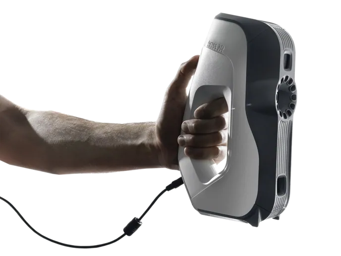
Artec Eva
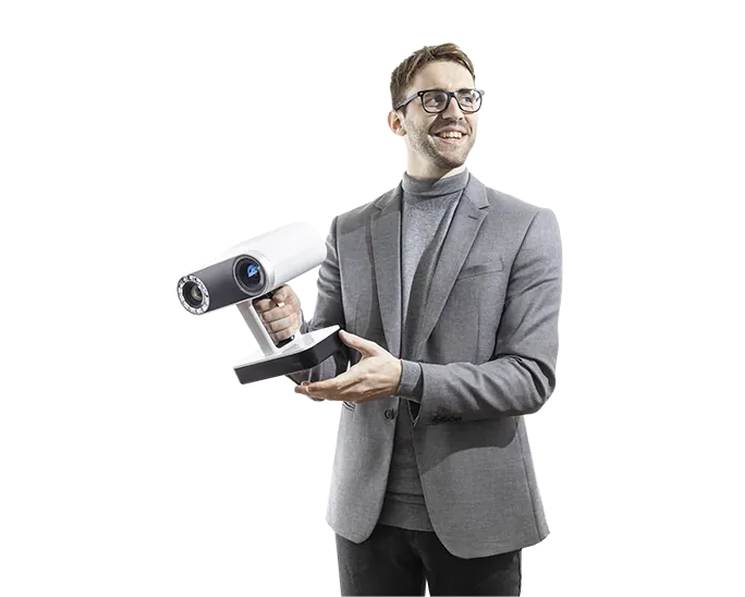
A handheld structured-light scanner that records a whole body in colour in one session, so the external findings sit in a measurable 3D model alongside the CT. For a single wound or a bone defect, Artec Spider II adds finer detail at up to 0.05 mm.
An examination of a body after death that uses imaging instead of dissection: post-mortem CT, often MRI and CT angiography, 3D scanning of the body surface and, where needed, needle biopsies. It is also called a virtopsy.
What is the difference between a virtual autopsy and a digital autopsy?
Mostly the name. Virtopsy comes from the Swiss research project that developed the method; digital autopsy is the name used for CT-based post-mortem services in the UK. Both rely on post-mortem CT; a full virtopsy adds surface scanning, angiography and image-guided biopsy.
Can a virtual autopsy replace a traditional autopsy?
For many sudden natural deaths, CT with coronary angiography can avoid an invasive autopsy, according to a 2017 Lancet study, but imaging still misses some conditions, such as pulmonary embolism. The same researchers recommend using both as the reference standard.
What are the disadvantages of virtual autopsy?
It misses some natural disease that dissection finds, cannot test tissue without a needle sample, needs specialist equipment and radiologists, and does not record the colour and detail of the skin without photographs or a surface scan.
Who performs a virtual autopsy?
Radiologists and forensic pathologists together. The radiologist reads the scans; the pathologist relates the findings to the circumstances of the death and, with the coroner or medical examiner, decides whether an autopsy is still needed.
Why do some families ask for a virtual autopsy?
Because it examines the body without opening it. Where a family's faith or wishes rule out an invasive autopsy, imaging can still establish the cause of death in many cases.
Tell us what you need to record at autopsy, from whole bodies to individual wounds and bone defects, and see how a scan captures it and what you can still measure afterwards.